אנו משתמשים ב-Cookies כדי לשפר את החוויה שלך. כדי לקיים ההנחיה החדשה של e-Privacy, עלינו לבקש את הסכמתך להגדיר את ה-Cookies. קבלת מידע נוסף.

מחיר מבצע
327.00 ₪
מחיר רגיל
392.00 ₪
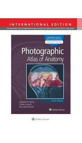
Photographic Atlas of Anatomy, International Edition 9e
מחיר מבצע
327.00 ₪
מחיר רגיל
392.00 ₪
ISBN13
9781975151560
מהדורה
9th Ed
הוצאה לאור
Wolters Kluwer Health
זמן אספקה
במלאי, (זמן אספקה 5 ימי עסקים)
עמודים / Pages
752
פורמט
Paperback
תאריך יציאה לאור
1 בפבר׳ 2021
Photographic Atlas of Anatomy features outstanding full-color photographs of actual cadaver dissections, with accompanying schematic drawings and diagnostic images, to help students develop an unparalleled mastery of human anatomy with ease. Depicting anatomic structures more realistically than illustrations in traditional atlases, this proven resource shows students exactly what they will see in the dissection lab. Chapters are organized by region in the order of a typical dissection, with each chapter presenting regional anatomical structures in a systematic manner.
This updated 9th edition includes revised content throughout and features additional cadaver dissection photos, medical imaging, and clinical illustrations, as well as a new appendix with learning resources that strengthen students’ understanding of the vascular, lymphatic, muscular, and nervous systems.
- UPDATED! Chapters organized by region guide you through the order of a typical dissection.
- NEW! Appendix with learning resources reinforces your understanding of the vascular, lymphatic, muscular, and nervous systems.
- More than 1,200 full-color dissection photos, medical imaging, and clinical illustrations —all new or updated— depict key anatomical distinctions and functional connections as seen in the dissection lab.
- Authentic photographic reproduction of colors, structures, and spatial dimensions familiarize you with the human anatomy as seen in the dissection lab and on the operating table.
- Functional connections between single organs, the surrounding tissue, and organ systems are clarified to help you prepare for the dissection lab and practical exams.
- Dissections illustrate the regional anatomy in layers "from the outside in" to prepare you for the lab and operating room.
- Clinical comments strengthen your understanding and clinical readiness.
אטלס אנטומיה מקיף ומוערך המציג צילומי צבע באיכות גבוהה של דיסקציות אמיתיות לצד איורים סכמטיים והדמיות רפואיות. הגישה הצילומית מאפשרת לסטודנטים לראות את מבני גוף האדם כפי שהם מופיעים בפועל במעבדת האנטומיה ובחדר הניתוח, ולהבין טוב יותר את הקשרים בין האיברים, הרקמות ומערכות הגוף השונות.
המהדורה התשיעית כוללת מאות תמונות מעודכנות, חומר קליני נוסף ונספח לימודי חדש המסייע להבנת מערכות כלי הדם, הלימפה, השרירים ומערכת העצבים. זהו מקור לימוד חיוני לסטודנטים לרפואה, מקצועות הבריאות ואנשי צוות רפואי הזקוקים להבנה ויזואלית ומעמיקה של האנטומיה האנושית.


-
 The 3D Atlas of Trigger Pointsמחיר מבצע 420.00 ₪ מחיר רגיל 554.00 ₪
 -

-
-

-
 Gray's Anatomy For Students International Editionמחיר מבצע 210.00 ₪ מחיר רגיל 235.00 ₪






| מהדורה | 9th Ed |
|---|---|
| עמודים / Pages | 752 |
| פורמט | Paperback |
| הוצאה לאור | Wolters Kluwer Health |
| תאריך יציאה לאור | 1 בפבר׳ 2021 |
Login and Registration Form