אנו משתמשים ב-Cookies כדי לשפר את החוויה שלך. כדי לקיים ההנחיה החדשה של e-Privacy, עלינו לבקש את הסכמתך להגדיר את ה-Cookies. קבלת מידע נוסף.

1,856.00 ₪
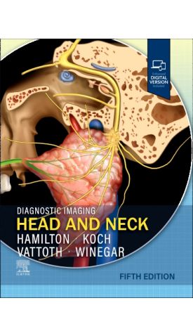
Diagnostic Imaging: Head And Neck
1,856.00 ₪
ISBN13
9780443378904
מהדורה
5th Edition
הוצאה לאור
Elsevier
זמן אספקה
21 ימי עסקים - לא במלאי בארץ
עמודים / Pages
1392
פורמט
Hardback + online
תאריך יציאה לאור
8 בדצמ׳ 2025
Covering the entire spectrum of this fast-changing field, the fifth edition of Diagnostic Imaging: Head and Neck is an invaluable resource for neuroradiologists, general radiologists, and trainees―anyone who requires an easily accessible, highly visual reference on today’s head and neck imaging. Drs. Bronwyn E. Hamilton, Bernadette L. Koch, Surjith Vattoth, Blair A. Winegar, and their team of highly regarded experts provide updated information on disease identification and imaging techniques to help you make informed decisions at the point of care. The layout is image rich, with succinct bulleted text that quickly conveys details, making it a useful learning tool as well as a handy reference for daily practice.
Key Features
- Serves as a one-stop resource for key concepts and information on head and neck imaging, including disease identification, imaging techniques, and details on tumor staging and classification.
- Reflects recent updates in genetic and molecular characterization of tumors, congenital malformations, and inflammatory/autoimmune disorders, which all have implications for targeted precision therapies.
- Offers 400+ concise, informative chapters, with content divided into sections based on head and neck spaces and anatomic regions, as well as on cancers, posttreatment appearances, pediatric lesions, and syndromic diseases.
- Features 2,800+ print images (with another 4,500+ additional images in the complimentary eBook), including radiologic images, full-color medical illustrations, clinical and gross pathologic photographs, and histologic images.
- Provides new and expanded content on genomic characterization of head and neck cancers; new characterizations of sinonasal tract and skull base tumors through distinctive genetic and molecular signatures; newer imaging techniques for neuroendocrine tumors and metastatic spread; whole-body imaging surveillance strategies for hereditary tumor syndromes; and much more.
- Emphasizes multidisciplinary involvement to help radiologists navigate the intersection of multiple specialties and confidently guide clinicians and surgeons toward the most appropriate diagnosis and treatment.
- Includes a complimentary eBook with additional images, text, and references, plus search, customization, note-taking, highlighting, and read-aloud functionality.
המהדורה החמישית של Diagnostic Imaging: Head and Neck מהווה מקור ידע מקיף ועדכני לאנשי רדיולוגיה, נוירורדיולוגיה ומתמחים העוסקים בדימות ראש וצוואר. הספר משלב אלפי תמונות איכותיות, איורים רפואיים וטקסט תמציתי המאפשר גישה מהירה למידע חיוני לצורך אבחון וקבלת החלטות קליניות.
הספר סוקר את כל תחומי הדימות של הראש והצוואר, כולל זיהוי מחלות, טכניקות הדמיה מתקדמות, דירוג וסיווג גידולים, מומים מולדים, מחלות דלקתיות ואוטואימוניות, וכן התפתחויות עדכניות בתחום הגנטיקה והמולקולריה המשפיעות על טיפולים מותאמים אישית.
התוכן מחולק ליותר מ־400 פרקים קצרים וממוקדים, המסודרים לפי אזורים אנטומיים ומצבים קליניים שונים, לרבות גידולים, ממצאים לאחר טיפול, מחלות בילדים ותסמונות נדירות. הספר כולל למעלה מ־2,800 תמונות בדפוס ואלפי תמונות נוספות בגרסה הדיגיטלית.
זהו מדריך מקצועי מעשי המיועד לשימוש יומיומי, המסייע לרדיולוגים ולצוותים רב־תחומיים להגיע לאבחנה מדויקת ולבחירת הטיפול המתאים במגוון רחב של מצבים רפואיים.

-

-
 Grainger & Allison's Diagnostic Radiologyמחיר מבצע 1,765.00 ₪ מחיר רגיל 2,273.00 ₪



| מהדורה | 5th Edition |
|---|---|
| עמודים / Pages | 1392 |
| פורמט | Hardback + online |
| הוצאה לאור | Elsevier |
| תאריך יציאה לאור | 8 בדצמ׳ 2025 |
| תוכן עניינים |
|
| Author | Bronwyn E. Hamilton, Bernadette L. Koch, Surjith Vattoth, Blair A. Winegar |
Login and Registration Form