אנו משתמשים ב-Cookies כדי לשפר את החוויה שלך. כדי לקיים ההנחיה החדשה של e-Privacy, עלינו לבקש את הסכמתך להגדיר את ה-Cookies. קבלת מידע נוסף.
1,359.00 ₪
Medaka: Biology, Management, and Experimental Prot ocols
1,359.00 ₪
ISBN13
9781119575290
יצא לאור ב
Hoboken
מהדורה
2nd Edition
זמן אספקה
21 ימי עסקים
עמודים
360
פורמט
Hardback
תאריך יציאה לאור
25 באוק׳ 2019
מחליף את פריט
1A813808710
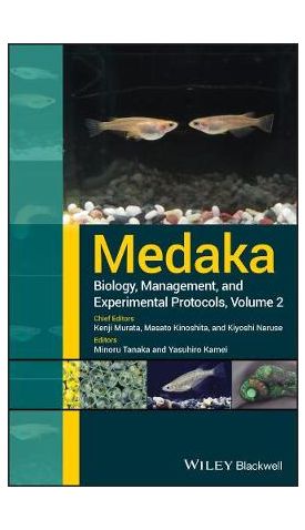

Medaka, Biology, Management, and Experimental Protocols, Volume 2 is the second in the series, with volume 1 published in 2009. The purpose of Volume 2, is to familiarize scientist worldwide with the advantages of using medaka inexperimental designs, to facilitate research using medaka, and to stimulate progress in research by adopting medaka as a model animal.
In Volume 2, the authors provide additional information and current protocols that have been recently developed, or modified, to successfully raise medaka fish under stable culture conditions in the laboratory; and how to use medaka as a model animal. This volume describes new technologies developed after 2009, using the fish as a molecular tool in the fields of life science, evolution, ecology and toxicology. It provides an informational bridge that spans the varied research disciplines and abilities that range from undergraduate education through the level of senior researcher, and addresses the value to science of medaka's adoption as a model animal.
Written by experts and pioneers in the use of medaka as the model animal in their scientific fields. The authors describe their experimental protocols in detail and the rationale for the chosen protocols in achieving their conceptual goals. The editors recommend that users read the previous procedures of Volume 1 that describe the maintenance of medaka; and use this information to create or modify the current fish-maintenance systems to improve and advance the science and
technology.
Medaka, Biology, Management, and Experimental Protocols, Volume 2 format is designed to capture the thoughts and methods of researchers that use medaka as a model animal; and to make this expertise accessible to students, beginning researchers and senior researchers, who might become intrigued with using medaka fish as the model animal in their own works.
To accomplish this, and following a reading of Volume 1, the reader is provided step-by step specifics for each protocol that allows application of the fish in their own work. The information includes specific information to facilitate ease of adoption: minute details such as reagents used in methodology, instrumentation, and other essential requirements. It is anticipated that this highly practical format will encourage the reader to use medaka as a model animal; and permit the reader to bring new concepts into personal practice in a more efficient manner.
| מהדורה | 2nd Edition |
|---|---|
| עמודים | 360 |
| מחליף את פריט | 1A813808710 |
| פורמט | Hardback |
| ISBN10 | 111957529X |
| יצא לאור ב | Hoboken |
| תאריך יציאה לאור | 25 באוק׳ 2019 |
| תוכן עניינים | Chapter 1: Medaka Management 1.0 Introduction 1.1 Targetable nuclease system for genome editing 1.1.1 Outline of medaka life-cycle in the wild 1.1.2 Preparation of normal rearing conditions of medaka in the laboratory and procedures for breeding 1.2 Standardized culture and growth curve 1.2.1 Characteristics and selection of strains 1.2.2 Management of medaka eggs and fish 1.2.3 Maintenance of breeding tanks during breeding 1.2.4 Anesthesia References Chapter 2: Medaka and Oryzias species as model organisms and the current status of medaka biological resources 2.0 Abstract 2.1 Common and unique futures of medaka and related species as model organisms 2.2 Phylogenetic relationships of medaka and the related species 2.2.1 The Javanicus Species group 2.2.2 The latipes species group 2.2.3 The celebensis species group 2.3 BAC resources of species related to Medaka 2.4 National BioResource Project Medaka (NBRP Medaka) 2.4.1 Support for visiting researchers 2.5 Acknowledgments References Chapter 3: Looking at Adult Medaka 3.1 General Morphology 3.1.1 Secondary sexual characters 3.1.2 Body color 3.2 Anatomy and Histology 3.2.1 Observations of internal organs 3.2.2 Horizontal and sagittal sections of juvenile medaka 3.2.3 Nervous system 3.2.4 Endocrine system 3.2.5 Gonads 3.2.6 Kidney Column 3. How to make sections of a meture ovary for histological analysis References Chapter 4: Looking at Medaka Embryos 4.1 Development of Various Tissues and Organs 4.1.1 Developmental stages 4.1.2 Brain 4.1.3 Hatching gland 4.1.4 Eye development 4.1.5 Branchial arch and jaws 4.1.6 Vasculature 4.1.7 Blood cells (hematopoiesis) 4.1.8 Heart 4.1.9 Kidney 4.1.10 Thymus 4.1.11 Gut and liver 4.1.12 Bones 4.1.13 Fins 4.1.14 Gonads 4.2 Medaka EGG Envelope and Hatching Enzyme 4.2.1 Overview 4.2.2 Preparation of a hatching enzyme solution from hatching liquid 4.2.3 Simple method for preparing hatching enzyme solution 4.2.4 Solubilization of the egg envelope using hatching enzyme 4.3 Observation of Embryos (Embedding Embryos) 4.3.1 Anesthesia of Embryos using MS-222 4.3.2 Observation of embryos (mounting) 4.4 Whole Mount in situ Hybridization (see Section 4.1.8 for a similar protocol) 4.4.1 Fixation and storage 4.4.2 Rehydration, proteinase K Treatment and post-fixation at RT 4.4.3 Hybridization and washing 4.4.4 Immunoreaction and washing antibodies 4.4.5 Staining 4.5 Embedding in a Plastic Resin (Technovit 7100) 4.5.1 Agarose mounting (Figure 4.48) 4.5.2 Dehydration and infiltration (Figure 4.48) 4.5.3 Polymerization (Figure 4.48) Column 4.3 Pigment cells (Figure 4.49) Column 4.4 Kupffer's vesicle References Chapter 5: Reproductive Behavior of Wild Japanese Medaka 5.1 Wild Japanese medaka 5.2 Reproductive behavior of wild medaka 5.2.1 Aggressive behavior 5.2.2 Spawning behavior 5.2.3 Egg deposition behavior 5.2.4 Egg discarding behavior 5.2.5 School and aggregation 5.3 Conclution References Chapter 6: Cryopreservation and Transplantation of Medaka Germ Cells 6.1 Introduction 6.2 Cryopreservation of medaka testes 6.2.1 Solutions 6.2.2 Materials 6.2.3 Procedures 6.3 Transplantation of thawed testicular cells into recipient larvae 6.3.1 Solutions 6.3.2 Materials 6.3.3 Procedures Column 6. Production of triploid medaka References Chapter 7: Genome Editing 7.0 Introduction 7 1 Outline of targeted genome editing using nucleases 7.2 Preparation of CRISPR/Cas9 genome editing tools 7.2.1 Materials 7.2.2 Procedure for production of custom-designed sgRNA 7.2.3 Procedure for production of capped RNA encoding a Cas9 nuclease 7.3 Preparation of custom-designed TALENs 7.3.1 Materials 7.3.2 Procedure for preparation of the TALEN assembly system 7.3.3 Procedure for design and construction of custom-designed TALENs 7.4 Heteroduplex mobility assay - A simple method to detect targeted genome modification 7.5 How to establish gene KO line 7.6 How to establish gene konckin strains Column 7. Utilization of crRNA, tracrRNA, Cas9 protein Appendix 7 Simple genomic DNA preparation by an alkaline lysis method References Chapter 8: Photo-inducible gene expression in medaka 8.0 Outline of IR-LEGO 8.1 Practical strategies of IR-LEGO in medaka study 8.2 Laser irradiation conditions and sample preparation 8.3 Caution in maintaining strains 8.4 Other use of IR-LEGO 8.5 Summary and the future References Chapter 9: Screening and Testing Methods of Endocrine Disrupting Chemicals using Medaka 9.1 Applied Toxicity Tests for Endocrine Disruptors 9.2 Detection of androgenic and anti-androgenic chemicals using medaka 9.2.1 The formation of "papillary processes" on anal fin rays as an indicative phenotype for exposure of androgenic and/or anti-androgenic chemicals 9.2.2 Candidate biomarkers for assessing the action of androgenic and anti-androgenic chemicals 9.2.3 Visualization of androgenic and anti-androgenic activity as green fluorescence with spiggin-GFP (green florescence protein) medaka References Chapter 10: Application of the seawater medaka Oryzias melastigma (McClelland) for marine ecotoxicology 10.1 Background and Development of Oryzias melastigma for Marine Ecotoxicology 10.2 Marine Medaka Developmental Staging 10.3 Standard Breeding and Rearing Conditions 10.3.1 Seawater 10.3.2 Temperature 10.3.3 Photoperiod 10.3.4 Feeding 10.3.5 Embryo collection and rearing 10.3.6 Hatching and larvae collection 10.3.7 Larvae rearing 10.3.8 Larvae feeding 10.4 Raising Marine Medaka for Experimental Use 10.4.1 Experiments using adult fish 10.4.2 Experiments using larvae 10.5 Troubleshooting 10.5.1 Mass mortality 10.5.2 Low egg production 10.5.3 Extensive algal growth 10.6 How to Obtain Marine Medaka O. melastigma 10.7 Experimental Protocols Using Marine Medaka 10.8 Immunotoxicity Assessment: the Bacteria Challenge Assays 10.8.1 SOP for adult bacterial challenge assay 10.8.2 SOP for larval bacterial challenge assay 10.8.3 Age selection for larval bacterial challenge 10.9 Fish Dissection and the Whole Adult Histoarray 10.9.1 SOP for fish dissection 10.9.2 SOP for adult medaka histoarray 10.10 Embryo Chip 10.10.1 SOP for embryo and larvae histoarray Appendix 10 Materials for SOP for adult medaka histoarray References Chapter 11: Telomerase and Telomere Biology in Medaka 11.1 Background 11.2 SOP for quantification of telomerase activity using the Real-Time Quantitative Telomeric Repeat Amplification Protocol (RTQ-TRAP) 11.3 SOP for quantification of telomere length using Southern blotting analysis 11.4 SOP for quantification of telomere length using fluorescence in situ hybridization (q-FISH) References Chapter 12: Assessments of Medaka Skeletal Toxicity 12.1 Introduction 12.2 Methods 12.2.1 Embryonic exposures- Dioxin 12.2.2 Embryonic exposure- Dithiocarbamates 12.2.3 Whole-mount Alcian blue staining of hatchling/larvae (1) 12.2.4 Whole-mount Alizarin red staining of hatchlings/larvae 12.2.5 In vivo Alizarin complexone fluorescent staining for mineralized bone matrix 12.2.6 In vivo Calcein fluorescent staining for mineralized bone matrix 12.2.7 Confocal imaging of embryo/hatchling medaka 12.2.8 Morphological assessments 12.3 Results and discussion References Appendix: Solutions Index |
| זמן אספקה | 21 ימי עסקים |
Login and Registration Form